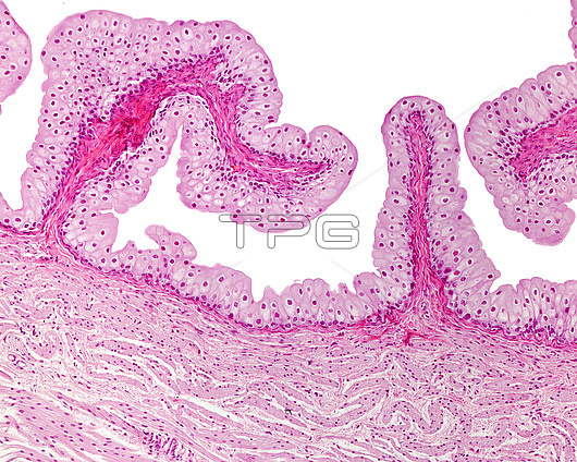
Urinary bladder, light micrograph. When empty, the luminal surface of the urinary bladder has numerous folds in its mucous layer. These folds are seen here. They consist of an inner connective tissue layer, the lamina propria, and a lining of urinary epithelium, or urothelium, a special type of transitional epithelium that lines both the ureter and the urinary bladder. Surrounding the lamina propria is a muscular layer, with smooth muscle myocyte bundles in oblique section and, at lower left, in longitudinal section.
| px | px | dpi | = | cm | x | cm | = | MB |
Details
Creative#:
TOP27562944
Source:
達志影像
Authorization Type:
RM
Release Information:
須由TPG 完整授權
Model Release:
N/A
Property Release:
N/A
Right to Privacy:
No
Same folder images:

 Loading