
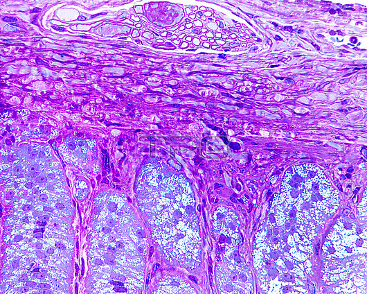
Outer limit of the adrenal gland, light micrograph. The upper half of the image corresponds to the adrenal capsule, in which a small bundle of myelinated fibres can be seen, probably destined for the medulla. Below is the zona glomerulosa formed by elongated cells that are arranged in palisades perpendicular to the capsular surface, forming elongated ovoid nests. These cells have lipid droplets as is typical of steroid-secreting cells, although as can be seen here, the droplets are smaller and more numerous than in the zona fasciculata. 0.5 micrometre semi-fine cut of material embedded in plastic and stained with toluidine blue.
| px | px | dpi | = | cm | x | cm | = | MB |
Details
Creative#:
TOP27276685
Source:
達志影像
Authorization Type:
RM
Release Information:
須由TPG 完整授權
Model Release:
N/A
Property Release:
N/A
Right to Privacy:
No
Same folder images:
 Loading