
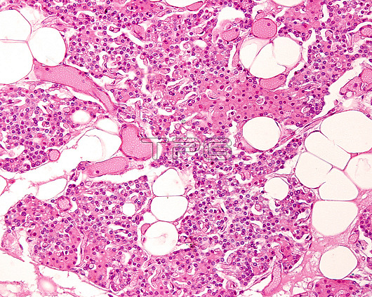
Light micrograph section of adult parathyroid gland stained with haematoxylin-eosin. The glandular parenchyma is made up of thick cell cords in which two cell types can be distinguished. The chief cells are very small in size, with their nuclei very close to each other. These cells predominate in the lower half of the image. The second type are oxyphilic cells, larger in size and with eosinophilic cytoplasm, which predominate in the upper part of the image. There are connective tissue septa among the cords, showing abundant blood vessels and some adipocytes.
| px | px | dpi | = | cm | x | cm | = | MB |
Details
Creative#:
TOP27149359
Source:
達志影像
Authorization Type:
RM
Release Information:
須由TPG 完整授權
Model Release:
N/A
Property Release:
N/A
Right to Privacy:
No
Same folder images:
 Loading