
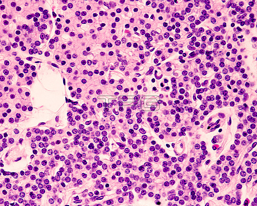
Light micrograph of the parathyroid gland of a young subject stained with haematoxylin-eosin. The glandular parenchyma is made up of thick cell cords in which two cell types can be distinguished. The chief cells are very small in size, leaving their nuclei very close to each other. These cells predominate in the lower half of the image. The second type are oxyphilic cells, larger in size, polygonal in shape and with eosinophilic cytoplasm, which can be seen in the upper left corner of the image.
| px | px | dpi | = | cm | x | cm | = | MB |
Details
Creative#:
TOP27149352
Source:
達志影像
Authorization Type:
RM
Release Information:
須由TPG 完整授權
Model Release:
N/A
Property Release:
N/A
Right to Privacy:
No
Same folder images:
 Loading