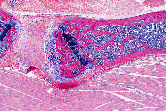
Light micrograph of a longitudinal section through the spine of a foetus showing developing vertebrae (tip of one at upper left, another across upper centre to upper right), the blocks of bone that make up the spine. Foetal bones are initially formed of cartilage (purple). Bone formation (ossification) starts from the middle of the cartilage and proceeds outwards. Areas where bone has already formed are red, areas of active ossification are dark purple. Pink and purple areas within the bone are bone marrow, a blood forming substance. An intervertebral disc (light pink), which acts as a shock absorber, can be seen between the vertebrae. Muscle tissue is across bottom. Magnification: x40 when printed at 15cm wide.
| px | px | dpi | = | cm | x | cm | = | MB |
Details
Creative#:
TOP27148546
Source:
達志影像
Authorization Type:
RM
Release Information:
須由TPG 完整授權
Model Release:
N/A
Property Release:
N/A
Right to Privacy:
No
Same folder images:

 Loading