
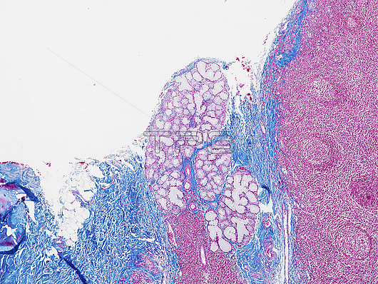
Light micrograph of a section through a nasopharyngeal tonsil, or adenoid (right) and a minor salivary gland (centre). The tonsils, part of the immune system, are large lymphatic nodules in a mucous membrane. The nasopharyngeal tonsils are located behind the nasal cavity. Each tonsil consists of lymphoid follicles (pink) that contain lymphocyte white blood cells. This minor salivary gland is a mixed gland with both mucus-producing cells (light pink) and serous-producing cells (darker pink). Connective tissue is blue. Magnification: x50 when printed at 15cm wide.
| px | px | dpi | = | cm | x | cm | = | MB |
Details
Creative#:
TOP27148524
Source:
達志影像
Authorization Type:
RM
Release Information:
須由TPG 完整授權
Model Release:
N/A
Property Release:
N/A
Right to Privacy:
No
Same folder images:
adenoidbiologicalbiologycellshealthyhistologicalhistologyimmunesystemimmunologicalimmunologylightmicrographlmlymphaticsystemlymphatictissuelymphocyteslymphoidnoduleslymphoidsystemmicroscopynasopharyngealtonsilno-onenobodynodenodulenonkeratinisednormalpharyngealtonsilsectionsectionedstratifiedsquamousepitheliumtissuetonsiltonsilswhitebloodcellsaciniglandulartubulelobulelobulesmixedmucousmucussalivasalivaryglandsecretoryserousminortubular
aciniadenoidbiologicalbiologybloodcellscellsepitheliumglandglandularhealthyhistologicalhistologyimmuneimmunologicalimmunologylightlmlobulelobuleslymphaticlymphaticlymphocyteslymphoidlymphoidmicrographmicroscopyminormixedmucousmucusnasopharyngealno-onenobodynodenodulenodulesnonkeratinisednormalpharyngealsalivasalivarysecretorysectionsectionedseroussquamousstratifiedsystemsystemsystemtissuetissuetonsiltonsiltonsiltonsilstubulartubulewhite
 Loading