
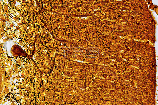
Light micrograph of a section through the cerebellum of the brain showing Purkinje nerve cells. Each Purkinje cell is composed of a flask-shaped cell body (one at left centre) from which branch numerous dendrites (brown strands). Purkinje cells are arranged at the junction of the molecular (right of Purkinje cell body) and granular (left of Purkinje cell body) layers of the cerebellum, which make up the grey matter of the brain. Nerve impulses flow to the Purkinje cells through their dendrites. The message is then passed on to the white matter deep in the cerebellar cortex. Nuclei (small dots) of other nerve cells, such as glial cells, can be seen throughout the grey matter. Magnification: x600 when printed at 15cm wide.
| px | px | dpi | = | cm | x | cm | = | MB |
Details
Creative#:
TOP27148126
Source:
達志影像
Authorization Type:
RM
Release Information:
須由TPG 完整授權
Model Release:
N/A
Property Release:
N/A
Right to Privacy:
No
Same folder images:
 Loading