
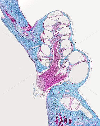
Light micrograph of a section through the fluid-filled canal of the cochlea of the inner ear. The canal is divided into three ducts (white). The vestibular membrane (thin diagonal lines) separates the upper vestibular duct from the central cochlear duct and the organ of Corti (thicker horizontal lines) lies between the cochlear duct and tympanic duct. Noise vibrations in the fluid in the canal cause vibrations in the vestibular membrane that in turn create vibrations in the organ of Corti. The tectorial membrane (very light blue at left of organ of Corti) vibrates and stimulates the sensitive hair cells on the basal membrane (blue at left of organ of Corti), which translate the vibrations to electrical signals that are sent to the brain via the auditory nerve (pink, centre). Magnification: x20 when printed at 15cm tall.
| px | px | dpi | = | cm | x | cm | = | MB |
Details
Creative#:
TOP27148057
Source:
達志影像
Authorization Type:
RM
Release Information:
須由TPG 完整授權
Model Release:
N/A
Property Release:
N/A
Right to Privacy:
No
Same folder images:
 Loading