
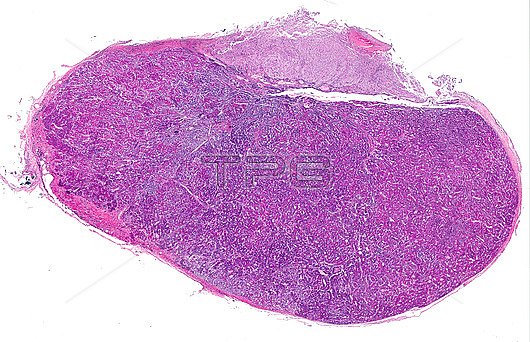
Light micrograph of a human pituitary. From bottom to top, the anterior lobe is highly stained due to its cellular richness, the intermediate lobe, poorly developed and showing cysts and, finally, the posterior lobe of the neurohypophysis, paler and with an appearance similar to that of nervous tissue. Surrounding the anterior lobe is a connective tissue capsule.
| px | px | dpi | = | cm | x | cm | = | MB |
Details
Creative#:
TOP26812153
Source:
達志影像
Authorization Type:
RM
Release Information:
須由TPG 完整授權
Model Release:
N/A
Property Release:
N/A
Right to Privacy:
No
Same folder images:
pituitaryglandadenohypophysisanatomyanteriorpituitaryglandbiologicalbiologyendocrineglandendocrinologyglandglandularhistologicalhistologyhormonehumantissuehumanbodybodyhypophysislightmicroscopemicroscopyneurohypophysisparsintermediaparsnervosasecretoryanatomicallightmicrographnobodyno-onebiologybiologicalhistologicalsection
adenohypophysisanatomicalanatomyanteriorbiologicalbiologicalbiologybiologybodybodyendocrineendocrinologyglandglandglandglandglandularhistologicalhistologicalhistologyhormonehumanhumanhypophysisintermedialightlightmicrographmicroscopemicroscopynervosaneurohypophysisno-onenobodyparsparspituitarypituitarysecretorysectiontissue
 Loading