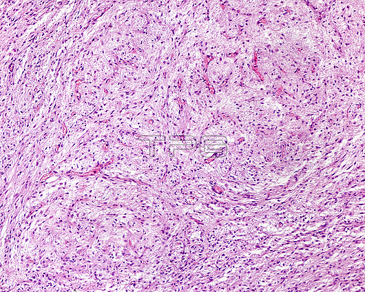
Light micrograph of the posterior lobe of the hypophysis (pituitary gland) stained with haematoxylin-eosin. It has numerous cells (pituicytes) separated by a fibrillar-like intercellular substance. In general, the appearance of the posterior lobe resembles that of a nervous tissue. Scattered throughout the tissue are blood vessels.
| px | px | dpi | = | cm | x | cm | = | MB |
Details
Creative#:
TOP26812147
Source:
達志影像
Authorization Type:
RM
Release Information:
須由TPG 完整授權
Model Release:
N/A
Property Release:
N/A
Right to Privacy:
No
Same folder images:

 Loading