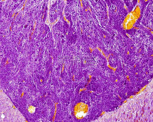
Light micrograph of a neurosecretion in the posterior pituitary (neurohypophysis) stained with Gabe's paraldehyde-fuchsin method. The neurosecretory material is distributed throughout the posterior lobe, showing few aggregates or Herring bodies. The orange-stained structures are blood vessels filled with red blood cells.
| px | px | dpi | = | cm | x | cm | = | MB |
Details
Creative#:
TOP26812134
Source:
達志影像
Authorization Type:
RM
Release Information:
須由TPG 完整授權
Model Release:
N/A
Property Release:
N/A
Right to Privacy:
No
Same folder images:
neurohypophysisanatomypituitaryglandbiologyendocrineglandendocrinologyglandglandularhistologicalhistologyhormonehumanbodyhypophysishypothalamuslightmicroscopemicroscopyparsnervosapituitaryglandpituitaryglandposteriorpituitaryglandsecretorylightmicrographhypothalamusgabefuchsinparaldehydenobodyno-onebiologybiologicalhistologicalsection
anatomybiologicalbiologybiologybodyendocrineendocrinologyfuchsingabeglandglandglandglandglandglandglandularhistologicalhistologicalhistologyhormonehumanhypophysishypothalamushypothalamuslightlightmicrographmicroscopemicroscopynervosaneurohypophysisno-onenobodyparaldehydeparspituitarypituitarypituitarypituitaryposteriorsecretorysection

 Loading