
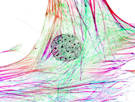
Inverted immunofluorescence micrograph of a murine fibroblast stained with an actin cytoskeleton probe (red/purple), anti-FGFR-1 (fibroblast growth factor receptor 1) antibody (blue/green), and nuclear DAPI (black). Colours were achieved with multiple z-stacks. Focal planes were achieved with an optical apotome.
| px | px | dpi | = | cm | x | cm | = | MB |
Details
Creative#:
TOP26739605
Source:
達志影像
Authorization Type:
RM
Release Information:
須由TPG 完整授權
Model Release:
N/A
Property Release:
N/A
Right to Privacy:
No
Same folder images:
cellcellsfibroblastnucleuscytoskeletonimmunofluorescencefluorescenceimmunofluorescentfluorescentlightmicrographbiologybiologicalnobodyno-onecellularstainstainedmurinemousemammalcytologycytologicalmicroscopyfibroblastgrowthfactorreceptor1FGFR-1FGFRdapinucleusactincytoskeletoncytoskeletalproteininverted
1FGFRFGFR-1actinbiologicalbiologycellcellscellularcytologicalcytologycytoskeletalcytoskeletoncytoskeletondapifactorfibroblastfibroblastfluorescencefluorescentgrowthimmunofluorescenceimmunofluorescentinvertedlightmammalmicrographmicroscopymousemurineno-onenobodynucleusnucleusproteinreceptorstainstained
 Loading