
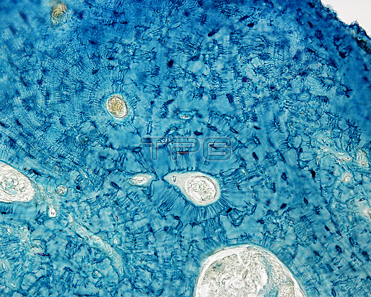
Light micrograph of compact bone stained with the Schmorl technique, showing osteocytes and their fine processes (blue). There are two osteons, each centred by a rounded space corresponding to the Haversian canal, which is occupied by a small blood vessel. The bone lamellae are arranged around them in concentric layers and the osteocytes are located between them. The numerous extensions that start from the osteocyte soma are striking and interconnect with each other, forming a complex labyrinth. The osteocytes closest to the centre of the osteon orient their processes towards the Haversian canal.
| px | px | dpi | = | cm | x | cm | = | MB |
Details
Creative#:
TOP26624868
Source:
達志影像
Authorization Type:
RM
Release Information:
須由TPG 完整授權
Model Release:
N/A
Property Release:
N/A
Right to Privacy:
No
Same folder images:
 Loading