
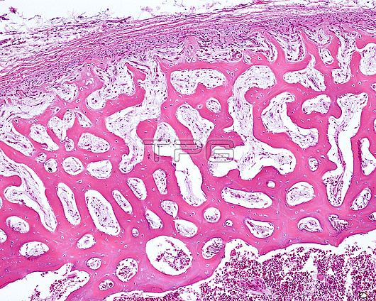
Peripheral area of the diaphysis of an embryonic bone, light micrograph. The periosteum is at right, showing two layers: an outer, or fibrous layer (pink), and an internal, or osteogenic layer, with a more cellular mesenchyme, where the osteoprogenitor cells are generated. In the more peripheral bone trabeculae, osteoid lines appear formed by osteoblasts that increases the thickness of the diaphyseal cortex. As can be seen in the image, the mean diameter of the trabeculae of immature bone tissue increases from the periosteum to the inside, since, once formed in the periosteum, new layers of bone matrix continue to be attached as time passes. In the internal part, resorption takes place and progressively augments the size of the medullary cavity.
| px | px | dpi | = | cm | x | cm | = | MB |
Details
Creative#:
TOP26624838
Source:
達志影像
Authorization Type:
RM
Release Information:
須由TPG 完整授權
Model Release:
N/A
Property Release:
N/A
Right to Privacy:
No
Same folder images:
 Loading