
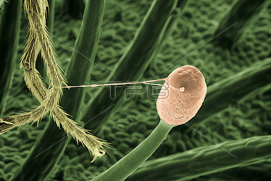
Scanning electron micrograph of a mucilage gland on a leaf of the cape sundew, Drosera capensis. The gland (orange) has been brushed by the leg of a small fly; the fly is caught by a thin thread of sticky mucilage, 10 microns in diameter. As it attempts to escape, the insect touches other glands, becoming trapped. The plant then uses secreted enzymes to break down the body of the insect, obtaining nutrients not available from the watery environment in which it grows. Analysis of the mucilage has shown it contains nanoparticles of diameter 50-70nm, that form fine fibres as the mucilage dries. These account for the very high elasticity of the mucilage, and also for its strength as an adhesive. A dried layer of the mucilage is also able to bind living cells strongly without harmful effect; this suggests a possible medical use for the material in wound healing and tissue engineering.
| px | px | dpi | = | cm | x | cm | = | MB |
Details
Creative#:
TOP26624798
Source:
達志影像
Authorization Type:
RM
Release Information:
須由TPG 完整授權
Model Release:
N/A
Property Release:
N/A
Right to Privacy:
No
Same folder images:
 Loading