
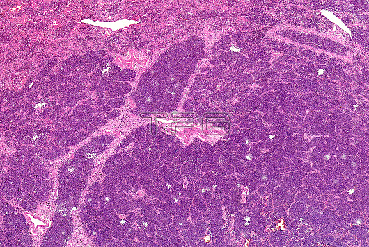
Light micrograph section through human lung tissue showing lobar pneumonia grey hepatisation. Grey hepatisation occurs 2 to 3 days following red hepatisation. The lung appears grey with liver-like consistency due to fibrinopurulent exudate, progressive disintegration of red blood cells and hemosiderin. This disease is also known as non-segmental pneumonia or focal non-segmental pneumonia. The most common causes are infections due to bacteria, such as, Streptococcus pneumoniae, Klebsiella pneumoniae and Legionella pneumophila.
| px | px | dpi | = | cm | x | cm | = | MB |
Details
Creative#:
TOP26387189
Source:
達志影像
Authorization Type:
RM
Release Information:
須由TPG 完整授權
Model Release:
N/A
Property Release:
N/A
Right to Privacy:
No
Same folder images:
anatomyanatomicalabnormalunhealthyhumananatomybiologybiologicalhumanpathologypathologyhistologyhistologicalhistopathologicalhealthcaremedicalunhealthylunglungsrespiratorysystemlungtissuelobarpneumoniagreyhepatisationredhepatisationliver-likeconsistencyfibropurulentexudateprogressivedisintegrationhemosiderinlungconditionlungdiseasenon-segmentalpneumoniafocalnon-segmentalpneumoniainfectionsbacterialinfectionsstreptococcuspneumoniaeKlebsiellapneumoniaelegionellapneumophilasectionillnessstainedpreparationvertebratevertebrateslightmicrographlightmicroscopelightmicroscopylmnobodyno-one
Klebsiellaabnormalanatomicalanatomyanatomybacterialbiologicalbiologyconditionconsistencydiseasedisintegrationexudatefibropurulentfocalgreyhealthcarehemosiderinhepatisationhepatisationhistologicalhistologyhistopathologicalhumanhumanillnessinfectionsinfectionslegionellalightlightlightliver-likelmlobarlunglunglunglunglungsmedicalmicrographmicroscopemicroscopyno-onenobodynon-segmentalnon-segmentalpathologypathologypneumoniapneumoniapneumoniapneumoniaepneumoniaepneumophilapreparationprogressiveredrespiratorysectionstainedstreptococcussystemtissueunhealthyunhealthyvertebratevertebrates
 Loading