
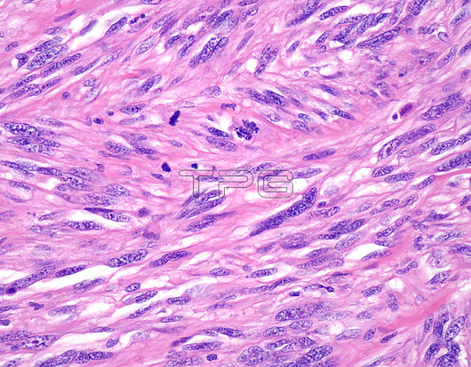
Spindle cell melanoma, light micrograph. Melanoma exhibits a variety of microscopic patterns. The tumour cells can be epithelioid, spindle shaped, signet ring type, or highly anaplastic with bizarre shape. This light micrograph shows spindle cell morphology that is commonly seen in melanomas arising in heel region (acral lentiginous melanoma).
| px | px | dpi | = | cm | x | cm | = | MB |
Details
Creative#:
TOP26298685
Source:
達志影像
Authorization Type:
RM
Release Information:
須由TPG 完整授權
Model Release:
N/A
Property Release:
N/A
Right to Privacy:
No
Same folder images:
ACTINICCANCERCANCEROUSCUTANEOUSDERMATOLOGYDERMATOLOGICALDERMATOPATHOLOGYDERMISEPIDERMISHISTOPATHOLOGICALHISTOPATHOLOGYLIGHTMICROGRAPHLIGHTMICROSCOPELIGHTMICROSCOPYMALIGNANCYMALIGNANTMALIGNANTMELANOMAMELANOCYTESMELANINMELANOMAMELANOMA-IN-SITUNEOPLASMONCOLOGYPATHOLOGICALPATHOLOGYPIGMENTPIGMENTATIONSKINCANCERSOLARRADIATIONSUNBURNSUN-DAMAGEDTUMOURSPINDLECELLLIGHTMICROGRAPHLMTUMOR
ACTINICCANCERCANCERCANCEROUSCELLCUTANEOUSDERMATOLOGICALDERMATOLOGYDERMATOPATHOLOGYDERMISEPIDERMISHISTOPATHOLOGICALHISTOPATHOLOGYLIGHTLIGHTLIGHTLIGHTLMMALIGNANCYMALIGNANTMALIGNANTMELANINMELANOCYTESMELANOMAMELANOMAMELANOMA-IN-SITUMICROGRAPHMICROGRAPHMICROSCOPEMICROSCOPYNEOPLASMONCOLOGYPATHOLOGICALPATHOLOGYPIGMENTPIGMENTATIONRADIATIONSKINSOLARSPINDLESUN-DAMAGEDSUNBURNTUMORTUMOUR
 Loading