
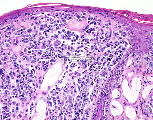
Nodular melanoma, light micrograph. Nodular melanoma (NM) accounts for 3-4% of all melanoma cases. It occurs in a relatively younger age group (5th or 6th decades) than other melanoma subtypes and is of shorter duration. It appears as a smooth firm elevated nodule or a polypoid mass covered by epidermis. The colour ranges from tan (amelanotic variants), brown, blue to black. The atypical melanocytes are arranged in infiltrating nests and show moderate cytologic atypia.
| px | px | dpi | = | cm | x | cm | = | MB |
Details
Creative#:
TOP26298676
Source:
達志影像
Authorization Type:
RM
Release Information:
須由TPG 完整授權
Model Release:
N/A
Property Release:
N/A
Right to Privacy:
No
Same folder images:
ACTINICCANCERCANCEROUSCUTANEOUSDERMATOLOGYDERMATOLOGICALDERMATOPATHOLOGYDERMISEPIDERMISHISTOPATHOLOGICALHISTOPATHOLOGYLIGHTMICROGRAPHLIGHTMICROSCOPELIGHTMICROSCOPYMALIGNANCYMALIGNANTMALIGNANTMELANOMAMELANOCYTESMELANINMELANOMAMELANOMA-IN-SITUNEOPLASMNODULARMELANOMAONCOLOGYPATHOLOGICALPATHOLOGYPIGMENTPIGMENTATIONSKINCANCERSOLARRADIATIONSUNBURNSUN-DAMAGEDTUMOURLIGHTMICROGRAPHLMTUMOR
ACTINICCANCERCANCERCANCEROUSCUTANEOUSDERMATOLOGICALDERMATOLOGYDERMATOPATHOLOGYDERMISEPIDERMISHISTOPATHOLOGICALHISTOPATHOLOGYLIGHTLIGHTLIGHTLIGHTLMMALIGNANCYMALIGNANTMALIGNANTMELANINMELANOCYTESMELANOMAMELANOMAMELANOMAMELANOMA-IN-SITUMICROGRAPHMICROGRAPHMICROSCOPEMICROSCOPYNEOPLASMNODULARONCOLOGYPATHOLOGICALPATHOLOGYPIGMENTPIGMENTATIONRADIATIONSKINSOLARSUN-DAMAGEDSUNBURNTUMORTUMOUR
 Loading