
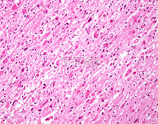
Diffuse astrocytoma, light micrograph. Infiltrating astrocytomas燼ccount for almost?0% of adult primary brain tumours. The usual location is燾erebral hemispheres燼nd the most commonly affected age group is?th to 6th decades. The histologic spectrum ranges from diffuse fibrillary astrocytomas (WHO Grade II; shown in this image), through anaplastic astrocytomas (WHO Grade III) to Glioblastoma (WHO Grade IV). WHO Grade I is reserved for Pilocytic astrocytomas. This image of diffuse astrocytoma (WHO Grade II) shows increased number of glial cells (cells with copious eosinophilic cytoplasm), mild nuclear pleomorphism, and a network of delicate astrocytic processes giving a fibrillary appearance to the background.
| px | px | dpi | = | cm | x | cm | = | MB |
Details
Creative#:
TOP25928914
Source:
達志影像
Authorization Type:
RM
Release Information:
須由TPG 完整授權
Model Release:
N/A
Property Release:
N/A
Right to Privacy:
No
Same folder images:
 Loading