
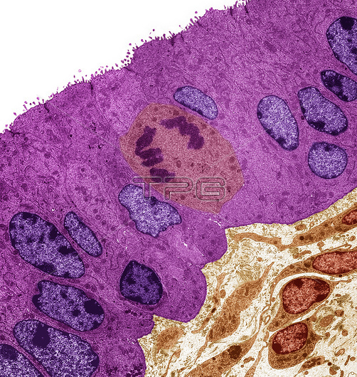
Fallopian tube, coloured transmission electron micrograph (TEM). Section through columnar epithelium from a fallopian tube during the secretory phase of the endometrial cycle when the ciliated cells are lacking. Large prominent nuclei are present (purple) with microvilli visible at the cell surface (top). A mitotic cell is seen center. Connective tissue is seen at the bottom right of the image. Magnification: x1000 when printed at 10 centimetres wide.
| px | px | dpi | = | cm | x | cm | = | MB |
Details
Creative#:
TOP25847293
Source:
達志影像
Authorization Type:
RM
Release Information:
須由TPG 完整授權
Model Release:
N/A
Property Release:
N/A
Right to Privacy:
No
Same folder images:
 Loading