
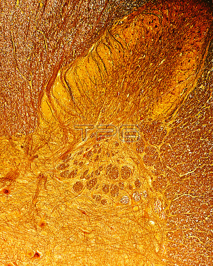
Spinal cord posterior horn, light micrograph. Posterior or dorsal horn of spinal cord showing the substantia gelatinosa of Rolando (lamina II) covering the nucleus propius (lamina III). In the base of dorsal horn, the reticular formation (a mixture of grey and white matter, exclusive to the cervical spinal cord) can be seen. The substantia gelatinosa of Rolando is crossed by dorsal column-medial lemniscus tract fibres. Cajal's silver nitrate method.
| px | px | dpi | = | cm | x | cm | = | MB |
Details
Creative#:
TOP25526359
Source:
達志影像
Authorization Type:
RM
Release Information:
須由TPG 完整授權
Model Release:
N/A
Property Release:
N/A
Right to Privacy:
No
Same folder images:
posteriorhornsubstantiagelatinosarolandocajalcellcellbodycentralnervoussystemcnsdendriteshistologylightmicroscopemicrographmicroscopemicroscopicmicroscopymultipolarnervoussystemnervoustissueneuroanatomyneurofibrilsneurohistologyneuroimagingneurologyneuronneurosciencesilvernitratesomaspinalcordtissuebiologyhistologyhistologicallightmicrographnobodyno-onelightmicrographlmneurologicalneuronebiological
biologicalbiologybodycajalcellcellcentralcnscorddendritesgelatinosahistologicalhistologyhistologyhornlightlightlightlmmicrographmicrographmicrographmicroscopemicroscopemicroscopicmicroscopymultipolarnervousnervousnervousneuroanatomyneurofibrilsneurohistologyneuroimagingneurologicalneurologyneuronneuroneneurosciencenitrateno-onenobodyposteriorrolandosilversomaspinalsubstantiasystemsystemtissuetissue
 Loading