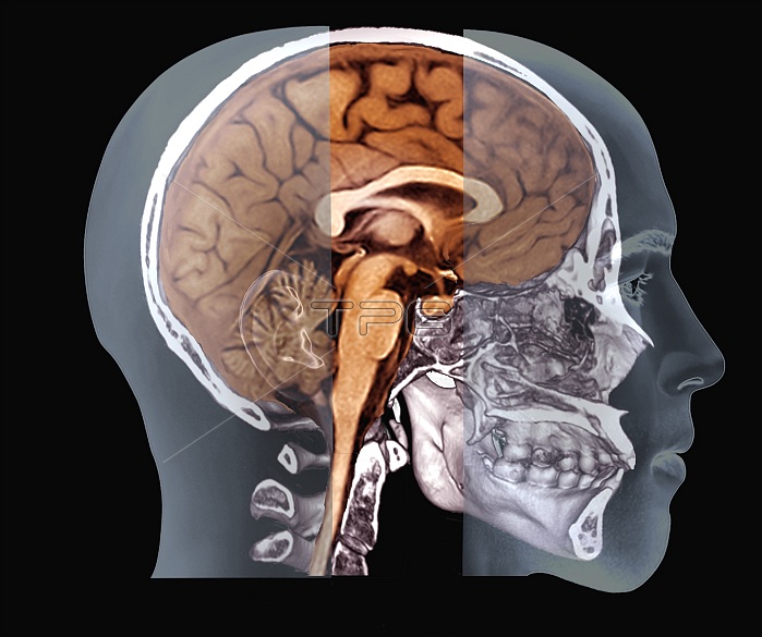
Human skull and brain. Combined coloured computed tomography (CT) and magnetic resonance imaging (MRI) 3D scans of a side view of the skull and brain of a 48-year-old man. The skull is shown by a CT scan, while the brain is shown with an MRI scan. Structures visible in the brain MRI scan include the cerebellum (rear base of the brain), the pituitary gland (behind the eyes), and the structures and ventricles between the two brain hemispheres (centre).
| px | px | dpi | = | cm | x | cm | = | MB |
Details
Creative#:
TOP25477136
Source:
達志影像
Authorization Type:
RM
Release Information:
須由TPG 完整授權
Model Release:
N/A
Property Release:
N/A
Right to Privacy:
No
Same folder images:
3DIMENSIONAL3-D3-DIMENSIONAL3DANATOMICALBIOLOGICALBONESCEREBELLUMCOMPUTEDTOMOGRAPHYCUTOUTCUTOUTSCUT-OUTCUT-OUTSCUTOUTCUTOUTSFALSE-COLOUREDFORTIESHEALTHYHYPOPHYSISLATERALMAGNETICRESONANCEIMAGINGMEDIANPLANENEUROLOGICALNO-ONENOBODYNORMALPITUITARYGLANDPROFILESAGITTALSIDEVIEWTHREEDIMENSIONALTHREE-DIMENSIONALVENTRICLESBONEHUMANBODYHEADANATOMYBIOLOGYNEUROLOGYADULT4840SMALEMANCOLOUREDCTSCANMRISCANSCANNER
33-D3-DIMENSIONAL3D40SMALE48ADULTANATOMICALANATOMYBIOLOGICALBIOLOGYBODYBONEHUMANBONESCEREBELLUMCOLOUREDCOMPUTEDCTCUTCUTCUT-OUTCUT-OUTSCUTOUTCUTOUTSDIMENSIONALDIMENSIONALFALSE-COLOUREDFORTIESGLANDHEADHEALTHYHYPOPHYSISIMAGINGLATERALMAGNETICMANMEDIANMRINEUROLOGICALNEUROLOGYNO-ONENOBODYNORMALOUTOUTSPITUITARYPLANEPROFILERESONANCESAGITTALSCANSCANSCANNERSIDETHREETHREE-DIMENSIONALTOMOGRAPHYVENTRICLESVIEW

 Loading