
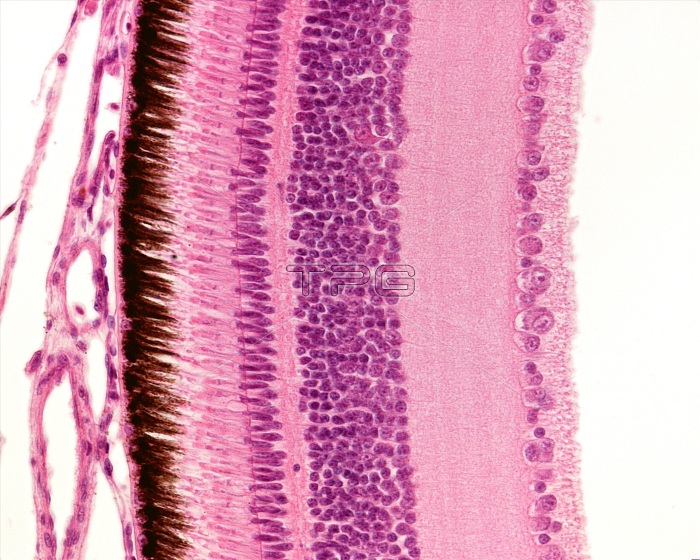
Light micrograph showing the layers of a bird's retina. From left to right, the retina layers are: pigment epithelium layer, rods and cones layer, outer nuclear layer, outer plexiform layer, inner nuclear layer, inner plexiform layer, ganglion cell layer and nerve fibre layer.
| px | px | dpi | = | cm | x | cm | = | MB |
Details
Creative#:
TOP25373477
Source:
達志影像
Authorization Type:
RM
Release Information:
須由TPG 完整授權
Model Release:
N/A
Property Release:
N/A
Right to Privacy:
No
Same folder images:
histologylightmicroscopenobodyno-onemicrographmicroscopemicroscopicmicroscopyhistologybiologyeyeeyeballophthalmologyophthalmicocularretinapigmentedepitheliumphotoreceptornervefibrelayerganglioncelllayerinnerplexiformlayerinnernuclearlayerouterplexiformlayerouternuclearlayerrodsconesbirdretinahistologicallightmicrographlmbiological
biologicalbiologybirdcellconesepitheliumeyeeyeballfibreganglionhistologicalhistologyhistologyinnerinnerlayerlayerlayerlayerlayerlayerlightlightlmmicrographmicrographmicroscopemicroscopemicroscopicmicroscopynerveno-onenobodynuclearnuclearocularophthalmicophthalmologyouterouterphotoreceptorpigmentedplexiformplexiformretinaretinarods
 Loading