
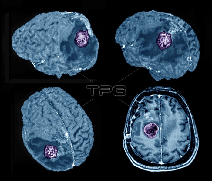
Recurrent glioma brain tumour. Coloured 3D and 2D magnetic resonance imaging (MRI) scans of the brain of a 60-year-old man who was operated on for a glioma brain tumour which has recurred (coloured mass). An axial scan is at lower right. This malignant tumour (cancer) arises from a type of glial (support) cell of the brain. The cancer is in the brain's left hemisphere and has returned 2 years after the initial operation. This cancer does not spread, but will damage the brain if not treated by surgery, radiotherapy or anti-cancer drugs.
| px | px | dpi | = | cm | x | cm | = | MB |
Details
Creative#:
TOP25179599
Source:
達志影像
Authorization Type:
RM
Release Information:
須由TPG 完整授權
Model Release:
N/A
Property Release:
N/A
Right to Privacy:
No
Same folder images:
3-D3DAXIALBLACKBACKGROUNDBRAINCANCERCANCEROUSCONDITIONCUTOUTCUTOUTSCUT-OUTCUT-OUTSCUTOUTCUTOUTSDIAGNOSISDIAGNOSTICSDISORDERFALSE-COLOUREDGLIALCELLSGLIOMALEFTHEMISPHEREMAGNETICRESONANCEIMAGINGMALIGNANTMEDICALNEURALNEUROLOGICALNO-ONENOBODYQUARTETSEQUENCESERIESSIXTIESTHREEDIMENSIONALTHREE-DIMENSIONALTUMORRECURRENTRECURRINGRETURNEDRECURRELAPSERELAPSEDDISEASECANCERTUMOURMALIGNANCYGROWTHHUMANBODYBRAINHEADMEDICINEONCOLOGYNEUROLOGYPATIENTADULT6060SMALEMANCOLOUREDMRISCANSCANNERFOUR4
3-D3D46060SMALEADULTAXIALBACKGROUNDBLACKBODYBRAINBRAINCANCERCANCERCANCEROUSCELLSCOLOUREDCONDITIONCUTCUTCUT-OUTCUT-OUTSCUTOUTCUTOUTSDIAGNOSISDIAGNOSTICSDIMENSIONALDISEASEDISORDERFALSE-COLOUREDFOURGLIALGLIOMAGROWTHHUMANHEADHEMISPHEREIMAGINGLEFTMAGNETICMALIGNANCYMALIGNANTMANMEDICALMEDICINEMRINEURALNEUROLOGICALNEUROLOGYNO-ONENOBODYONCOLOGYOUTOUTSPATIENTQUARTETRECURRECURRENTRECURRINGRELAPSERELAPSEDRESONANCERETURNEDSCANSCANNERSEQUENCESERIESSIXTIESTHREETHREE-DIMENSIONALTUMORTUMOUR
 Loading