
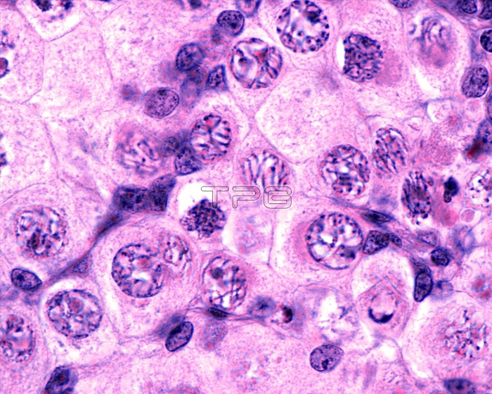
Light micrograph of a fetal ovary showing several oocytes (immature egg cells) undergoing meiosis (gamete formation). Three stages of prophase I are seen. On the left are three oocytes in leptotene, with chromosomes as thin strands. At centre and top, two oocytes are in zygotene with chromosomes in a bouquet. The other oocytes are in pachytene with chromosomes appearing as thick strands.
| px | px | dpi | = | cm | x | cm | = | MB |
Details
Creative#:
TOP24755137
Source:
達志影像
Authorization Type:
RM
Release Information:
須由TPG 完整授權
Model Release:
N/A
Property Release:
N/A
Right to Privacy:
No
Same folder images:
biologycellchromosomeseosinfemalefemalereproductivesystemgametogenesishaematoxylinhistologicalhistologymeiosismeioticgameteformationfoetalfoetusfetalfetusnobodyno-onelightmicrographmicroscopicmicroscopicanatomymicroscopynucleusoocyteoogoniaovarianovaryovogenesisprophasereproductivepachyteneleptotenezygotenebiological
anatomybiologicalbiologycellchromosomeseosinfemalefemalefetalfetusfoetalfoetusformationgametegametogenesishaematoxylinhistologicalhistologyleptotenelightmeiosismeioticmicrographmicroscopicmicroscopicmicroscopyno-onenobodynucleusoocyteoogoniaovarianovaryovogenesispachyteneprophasereproductivereproductivesystemzygotene
 Loading