
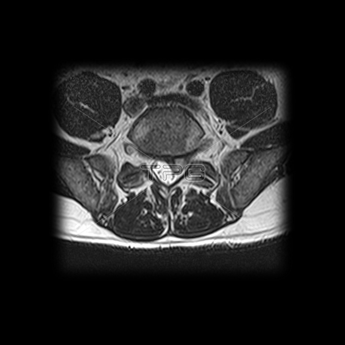
This axial (cross sectional) T2 weighted MRI image through the L5/S1 intervertebral disc level demonstrates a large left (on your right) sided herniation of the L5/S1 disc compressing and displacing the S1 nerve root posteriorly.
| px | px | dpi | = | cm | x | cm | = | MB |
Details
Creative#:
TOP22291093
Source:
達志影像
Authorization Type:
RM
Release Information:
須由TPG 完整授權
Model Release:
N/A
Property Release:
No
Right to Privacy:
No
Same folder images:
 Loading