
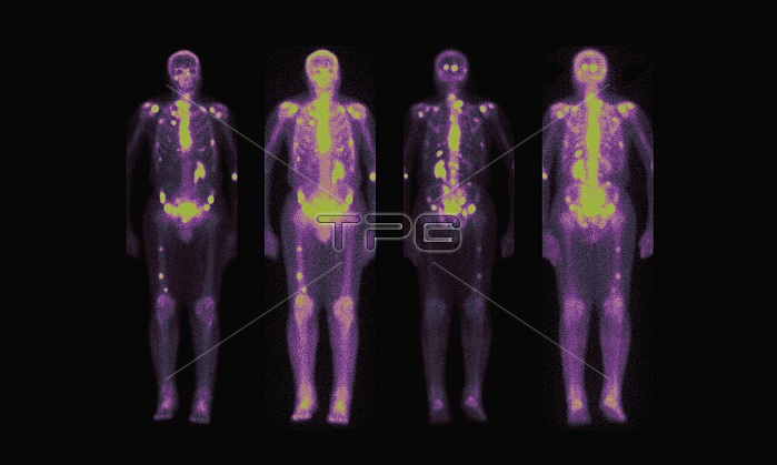
This composite of images is typical of what a nuclear medicine bone scan in a patient with spread of cancer to the skeleton looks like. This study is performed after a radio-isotope (nuclide) is injected into the blood stream. Several hours later a gamma camera (nuclear medicine camera) takes these images in different positions. The radio-isotope is picked up in bone where there is active turn-over (metabolism) of bony activity. In this instance the spread of lung cancer to the bones is shown as "hot spots" or areas of increased uptake of the nuclear agent (greenish yellow). The agent is excreted through the kidneys which allows us to see them. Note that this patient has only one functioning kidney. The non-functioning kidney does not contribute to the excretion of the radio-isotope and is therefore not seen.
| px | px | dpi | = | cm | x | cm | = | MB |
Details
Creative#:
TOP22289781
Source:
達志影像
Authorization Type:
RM
Release Information:
須由TPG 完整授權
Model Release:
N/A
Property Release:
No
Right to Privacy:
No
Same folder images:
 Loading