
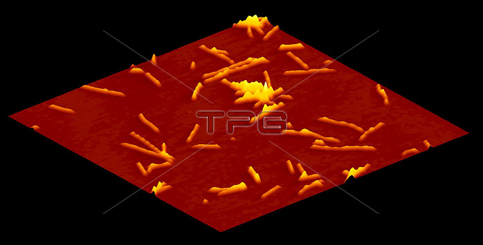
Atomic force micrograph (AFM) of amyloid fibrils, showing the topography (surface contours) of beta2- microglobulin (b2m) fibrils (yellow) on a mica surface (ultra-flat surface upon which the fibrils are imaged; red background). Fibrillar amyloid protein aggregates can be found in deposits or plaques associated with Alzheimer's disease, Creutzfeldt-Jakob disease (CJD) and diabetes. The fibrils were formed in vitro. Image is 10 x 10 micrometres. Supplied by Wei-Feng Xue, courtesy of Wellcome Images.
| px | px | dpi | = | cm | x | cm | = | MB |
Details
Creative#:
TOP22238949
Source:
達志影像
Authorization Type:
RM
Release Information:
須由TPG 完整授權
Model Release:
N/A
Property Release:
No
Right to Privacy:
No
Same folder images:
 Loading