
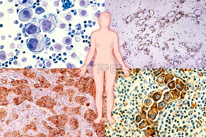
Illustration of the female body superimposed on a composite of micrographs of breast cancer cells. Clockwise from top left: (1) Human metastatic breast cancer in the pleural fluid. Stained with H&E and magnified to 400x. (2) Cross section of infiltrating ductal carcinoma of the breast with a small foci of breast cancer cells in which cd34 antibody has stained blood vessels and basement membrane. Magnification x100. (3) Human metastatic breast cancer in the lymph nodes. Stained by immunocytochemical for epithelial membrane antigen. Magnified to 400x. (4) An infiltrating ductal carcinoma of human breast origin is seen invading the breast tissue. The cytoplasm of the tumor cells is stained brown with a monoclonal antibody, which recognizes a carcinoembryonic type antigen (CEA) found within the malignant cells. Magnification is 313x.
| px | px | dpi | = | cm | x | cm | = | MB |
Details
Creative#:
TOP22233940
Source:
達志影像
Authorization Type:
RM
Release Information:
須由TPG 完整授權
Model Release:
N/A
Property Release:
No
Right to Privacy:
No
Same folder images:
 Loading