
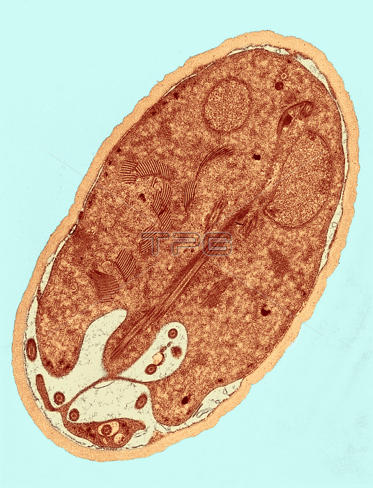
Color-enhanced Transmission Electron Micrograph (TEM) showing the cyst-stage of a Giardia sp. protozoan. The outer cyst wall is composed of filamentous and membranous portions, and is separated from the cytoplasm of the trophozoites contained within by the peritrophic space. This cyst wall is approximately 0.25 microns thick.
| px | px | dpi | = | cm | x | cm | = | MB |
Details
Creative#:
TOP22230701
Source:
達志影像
Authorization Type:
RM
Release Information:
須由TPG 完整授權
Model Release:
N/A
Property Release:
No
Right to Privacy:
No
Same folder images:
falsecolorcolorizationcolorizedcolorenhancementcolor-enhancedenhancementenhancedcystorganismmastigophorainfectiousinfectionsacromastigophoraprotozoanprotozoagiardiagiardiasisparasiticintestinaldiseasegastrointestinaldiseasedigestivesystemdiseaseemelectronmicrographtemtransmissionelectronmicrographhistopathologymicrographymicrographimagingconditiondiseasepathologyunhealthyabnormalmedicalscience
abnormalcolorcolorcolor-enhancedcolorizationcolorizedconditioncystdigestivediseasediseasediseasediseaseelectronelectronemenhancedenhancementenhancementfalsegastrointestinalgiardiagiardiasishistopathologyimaginginfectioninfectiousintestinalmastigophoramedicalmicrographmicrographmicrographmicrographyorganismparasiticpathologyprotozoaprotozoansacromastigophorasciencesystemtemtransmissionunhealthy
 Loading