
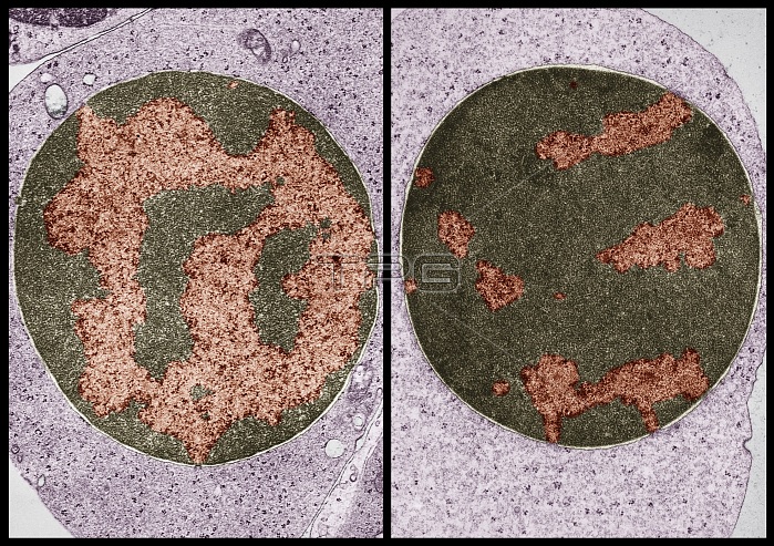
Transmission electron micrographs of a polychromatophilic erythroblast (left) and an orthochromatic erythroblast (right) from guinea pig bone marrow. On the left, cell specialization is marked by a coarser chromatin pattern, with large masses of chromatin (colored red) distributed throughout the nucleus. On the right, the erythroblast is at an advanced stage of nuclear condensation, which explains the scarcity of interchromosomal nucleoplasm. The right-hand image also shows a diminished number of polyribosomes.
| px | px | dpi | = | cm | x | cm | = | MB |
Details
Creative#:
TOP22230364
Source:
達志影像
Authorization Type:
RM
Release Information:
須由TPG 完整授權
Model Release:
N/A
Property Release:
No
Right to Privacy:
No
Same folder images:
 Loading