
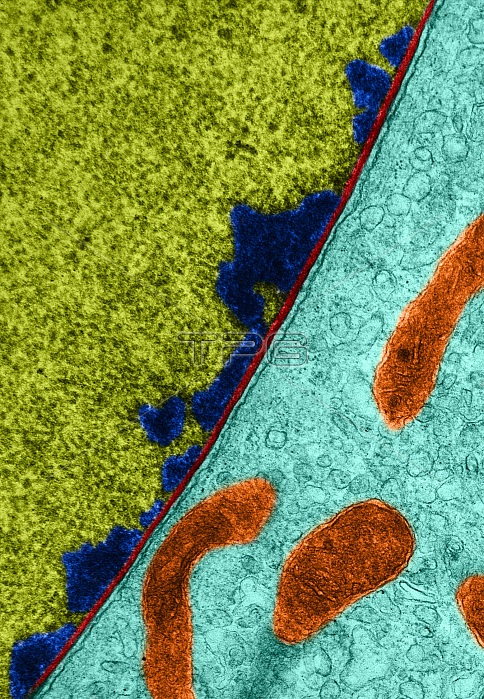
Transmission electron micrograph of an intestinal epithelial cell of Amphiuma tridactylum. The fibrous lamina (in red) is visible as a continuous dense layer about 60 nm thick interposed between the inner nuclear membrane (light blue) and the heterochromatin (dark blue).
| px | px | dpi | = | cm | x | cm | = | MB |
Details
Creative#:
TOP22227821
Source:
達志影像
Authorization Type:
RM
Release Information:
須由TPG 完整授權
Model Release:
N/A
Property Release:
No
Right to Privacy:
No
Same folder images:
 Loading