
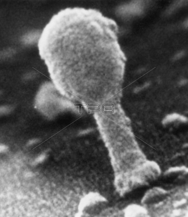
This untriggered T4 coliphage imaged by low-loss SEM shows the head, collar, tail, and end plate. Small projections arising from the end plate are similar in location and size to the spikes, which function in phage absorption. Rodlike structures are frequently observed extending from the end plate, or lying against the phage tail and projecting up onto the p hage head. These may be tail fibers which are partially obscured by the metal coating.
| px | px | dpi | = | cm | x | cm | = | MB |
Details
Creative#:
TOP22218629
Source:
達志影像
Authorization Type:
RM
Release Information:
須由TPG 完整授權
Model Release:
N/A
Property Release:
No
Right to Privacy:
No
Same folder images:
 Loading