
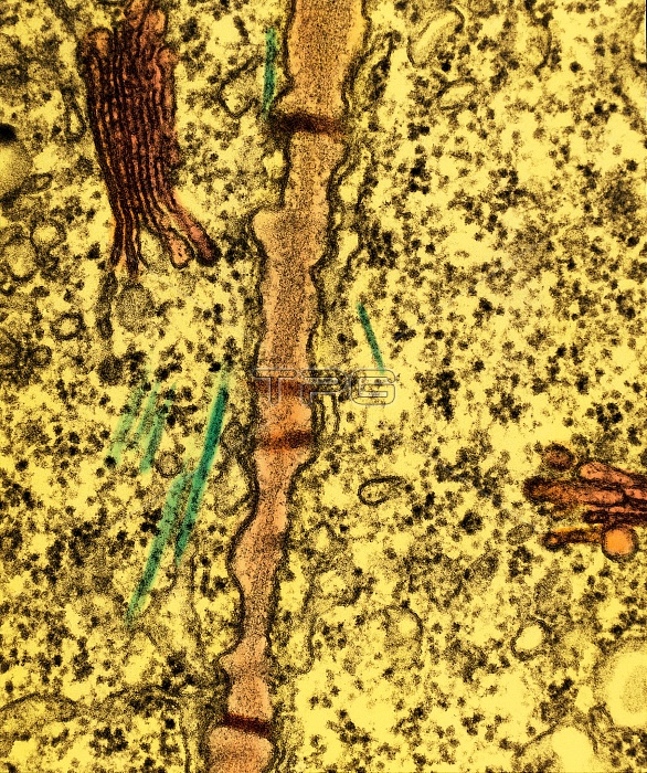
Color enhanced Transmission Electron Micrograph (TEM) section of cell wall (pink) traversed by plasmodesmata, plasmalemma-lined pores through which adjacent cells are linked by cytoplasmic strands; skeletal microtubules (green) lie next to the cell wall; the cytoplasm also contains Golgi bodies or dictyosomes (red), endoplasmic reticulum and many ribosomes. Magnification: 40,000x.
| px | px | dpi | = | cm | x | cm | = | MB |
Details
Creative#:
TOP22214043
Source:
達志影像
Authorization Type:
RM
Release Information:
須由TPG 完整授權
Model Release:
N/A
Property Release:
No
Right to Privacy:
No
Same folder images:
temtransmissionelectronmicrographelectronmicrographmicrographmicrographycolorizedcolorenhancedcolor-enhancedfalsecolorfalse-colorplantcellbiologybiologicalmicrobiologycellwallplasmodesmataplasmalemmaskeletalmicrotubulescytoplasmgolgibodiesdictysomesendoplasmicreticulumribosomeplantcellsfloraafricanvioletafricanvioletsvioletvioletsflowerflowerssaintpauliasaintpauliaionanthaionanthagolgibodygolgiapparatusgolgiapparatusesmicrotubulemicrotubulesribosomesplasmodesmatumrootroottiproottipsroottipcellroottipcellsbotanycellwallsplantcellwallplantcellwallss.ionantha
africanafricanapparatusapparatusesbiologicalbiologybodiesbodybotanycellcellcellcellcellcellcellscellscolorcolorcolor-enhancedcolorizedcytoplasmdictysomeselectronelectronendoplasmicenhancedfalsefalse-colorfloraflowerflowersgolgigolgigolgigolgiionanthaionanthaionanthamicrobiologymicrographmicrographmicrographmicrographymicrotubulemicrotubulesmicrotubulesplantplantplantplantplasmalemmaplasmodesmataplasmodesmatumreticulumribosomeribosomesrootrootrootrootroots.saintpauliasaintpauliaskeletaltemtiptiptiptipstransmissionvioletvioletvioletsvioletswallwallwallswalls
 Loading