
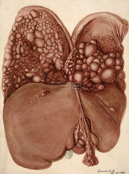
watercolor drawing of the liver and of the under surface of the diaphragm. The latter is almost covered by a number of rounded pedunculated growths, varying in size from that of a walnut to that of a pea. There are also a few on the liver, and several on the peritoneal surface of the falciform ligament of the liver. Other parts of the peritoneum, especially that covering the uterus and ovaries, were thickly studded with similar growths. Under the microscope these were found to be composed of encephaloid carcinoma, the glandular cells being large and spheroidal in shape. The seat of the primary disease was not determined. Leonard Mark, 1890.
| px | px | dpi | = | cm | x | cm | = | MB |
Details
Creative#:
TOP22167047
Source:
達志影像
Authorization Type:
RM
Release Information:
須由TPG 完整授權
Model Release:
N/A
Property Release:
No
Right to Privacy:
No
Same folder images:
 Loading