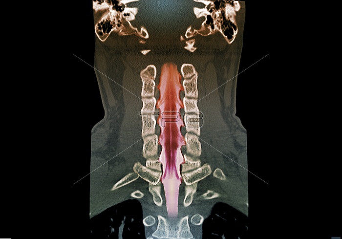
Neck bones and spinal cord. Coloured coronal computed tomography (CT) scan of the neck bones and spinal cord of a 43-year-old man. This scan uses a contrast medium to highlight the spinal cord and the nerves branching off it, in a technique known as myelography. The scan shows the normal anatomy of the cervical (neck) region of the spinal cord, the spinal nerve roots, and the anatomy of the cervical vertebrae (spinal bones).
| px | px | dpi | = | cm | x | cm | = | MB |
Details
Creative#:
TOP19633740
Source:
達志影像
Authorization Type:
RM
Release Information:
須由TPG 完整授權
Model Release:
N/A
Property Release:
N/A
Right to Privacy:
No
Same folder images:
ANATOMICALANTERIORBIOLOGICALBLACKBACKGROUNDBONESCENTRALNERVOUSSYSTEMCERVICALCNSCOMPUTEDTOMOGRAPHYCONTRASTMEDIUMCORONALFALSE-COLOUREDFORTIESFRONTALHEALTHYMYELOGRAMMYELOGRAPHYNEUROLOGICALNO-ONENOBODYNORMALSPINALVERTEBRAVERTEBRAEBONESPINALCORDNERVETISSUEHUMANBODYSPINEBACKBACKBONEVERTEBRALCOLUMNNECKANATOMYBIOLOGYNEUROLOGYADULT40S43MALEMANCTSCANCOLOUREDSCANNER
40S43MALEADULTANATOMICALANATOMYANTERIORBACKBACKBONEBACKGROUNDBIOLOGICALBIOLOGYBLACKBODYBONEBONESCENTRALCERVICALCNSCOLOUREDCOLUMNCOMPUTEDCONTRASTCORDCORONALCTFALSE-COLOUREDFORTIESFRONTALHEALTHYMANMEDIUMMYELOGRAMMYELOGRAPHYNECKNERVENERVOUSNEUROLOGICALNEUROLOGYNO-ONENOBODYNORMALSCANSCANNERSPINALSPINALSPINESYSTEMTISSUEHUMANTOMOGRAPHYVERTEBRAVERTEBRAEVERTEBRAL

 Loading