
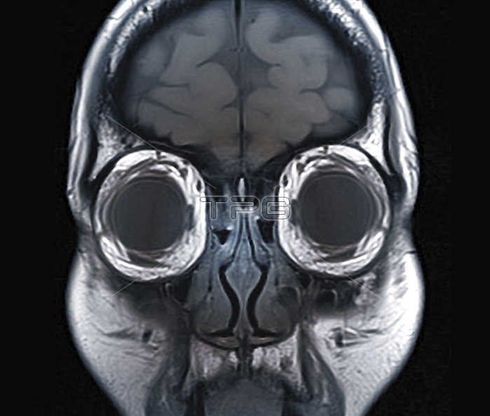
Eye anatomy and muscles. Coloured coronal magnetic resonance imaging (MRI) scan passing vertically through the head of a 33-year-old man, seen from the front, with the scan passing through the eyes. This scan shows some of the normal structures of the eyes and their support tissues, including the oblique muscles and the fat in the orbits of the eyes. Parts of the brain and nasal sinuses are also visible on this scan. This is a T1-weighted scan, with injection of gadolinium contrast medium.
| px | px | dpi | = | cm | x | cm | = | MB |
Details
Creative#:
TOP19633682
Source:
達志影像
Authorization Type:
RM
Release Information:
須由TPG 完整授權
Model Release:
N/A
Property Release:
N/A
Right to Privacy:
No
Same folder images:
ANATOMICALBIOLOGICALBLACKBACKGROUNDCONTRASTMEDIUMCORONALEYEORBITEYESFALSE-COLOUREDGADOLINIUMHEALTHYMAGNETICRESONANCEIMAGINGNASALNO-ONENOBODYNORMALOBLIQUEMUSCLESOCULAROPHTHALMICORBITALORBITALFATORBITSSECTIONSECTIONEDSINUSSINUSEST1RELAXATIONT1SCANT1WEIGHTEDTHIRTIESTISSUESORGANEYEMUSCLEBRAINTISSUEHUMANBODYHEADFACEBIOLOGYANATOMYOPHTHALMOLOGYADULT3330SMALEMANMRISCANCOLOUREDSCANNER
30SMALE33ADULTANATOMICALANATOMYBACKGROUNDBIOLOGICALBIOLOGYBLACKBODYBRAINCOLOUREDCONTRASTCORONALEYEEYEEYESFACEFALSE-COLOUREDFATGADOLINIUMHEADHEALTHYIMAGINGMAGNETICMANMEDIUMMRIMUSCLEMUSCLESNASALNO-ONENOBODYNORMALOBLIQUEOCULAROPHTHALMICOPHTHALMOLOGYORBITORBITALORBITALORBITSORGANRELAXATIONRESONANCESCANSCANSCANNERSECTIONSECTIONEDSINUSSINUSEST1T1T1THIRTIESTISSUEHUMANTISSUESWEIGHTED
 Loading