
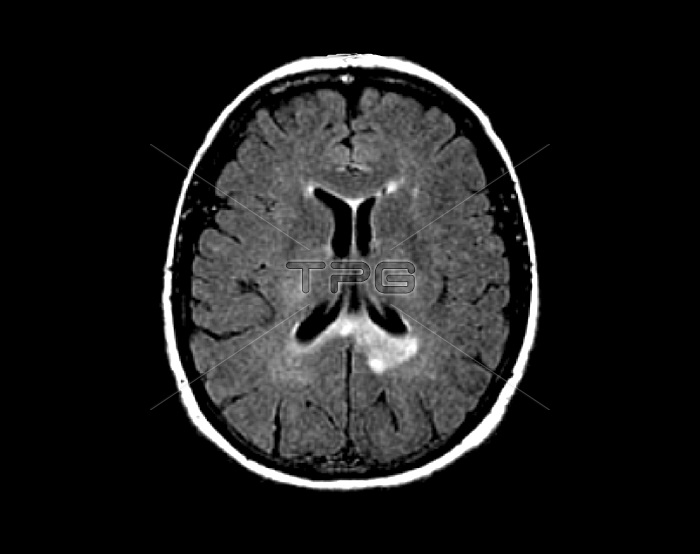
Multiple sclerosis. Axial magnetic resonance imaging (MRI) scan of the brain of a 42-year-old woman with multiple sclerosis. The scan shows white areas around the ventricles (centre) that are a sign of the demyelination of the nerves that occurs in this condition. This is a T1 MRI scan, with the use of gadolinium as a contrast medium.
| px | px | dpi | = | cm | x | cm | = | MB |
Details
Creative#:
TOP19633582
Source:
達志影像
Authorization Type:
RM
Release Information:
須由TPG 完整授權
Model Release:
N/A
Property Release:
N/A
Right to Privacy:
No
Same folder images:
ABNORMALAXIALBLACKBACKGROUNDCONDITIONCONTRASTMEDIUMCUTOUTCUTOUTSCUT-OUTCUT-OUTSCUTOUTCUTOUTSDEMYELINATEDDEMYELINATIONDIAGNOSISDIAGNOSTICSDISORDERFORTIESGADOLINIUMMAGNETICRESONANCEIMAGINGMEDICALNERVESNEURALNEUROLOGICALNO-ONENOBODYSECTIONSECTIONEDUNHEALTHYT1SCANT1WEIGHTEDSPINLATTICEDISEASESCLEROSISHUMANBODYBRAINMEDICINENEUROLOGYPATIENTADULT4240SFEMALEWOMANMRISCANSCANNERBLACK-AND-WHITEMONOCHROME
40SFEMALE42ABNORMALADULTAXIALBACKGROUNDBLACKBODYBRAINCONDITIONCONTRASTCUTCUTCUT-OUTCUT-OUTSCUTOUTCUTOUTSDEMYELINATEDDEMYELINATIONDIAGNOSISDIAGNOSTICSDISEASEDISORDERFORTIESGADOLINIUMIMAGINGLATTICEMAGNETICMEDICALMEDICINEMEDIUMMONOCHROMEMRINERVESNEURALNEUROLOGICALNEUROLOGYNO-ONENOBODYOUTOUTSPATIENTRESONANCESCANSCANSCANNERBLACK-AND-WHITESCLEROSISHUMANSECTIONSECTIONEDSPINT1T1UNHEALTHYWEIGHTEDWOMAN
 Loading