
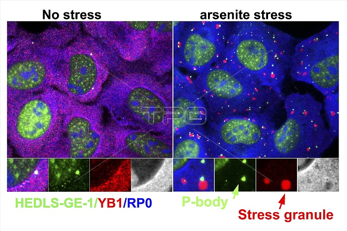
Arsenite-stressed cells. Immunofluorescence light micrograph of cells stained to show cell proteins and stress responses to arsenite. The image at left shows no stress, with the image at right showing stress due to arsenite (chemical compounds containing arsenic). The text at lower left gives the names of the proteins and the colours of their corresponding stains: HEDLS-GE-1 (green), YB1 (red, Y box binding protein 1), and RP0 (blue, ribosomal P0 protein). At right are inset images showing P-bodies (green, processing bodies containing RNA enzymes) and stress granules (red).
| px | px | dpi | = | cm | x | cm | = | MB |
Details
Creative#:
TOP16638044
Source:
達志影像
Authorization Type:
RM
Release Information:
須由TPG 完整授權
Model Release:
N/A
Property Release:
N/A
Right to Privacy:
No
Same folder images:
arsenicarsenitebiochemicalbiochemistrybiologicalbodycellcellbiologycellculturecellscellularcytologicalcytologycytoskeletonfluorescencefluorescenthedls-ge-1immunofluorescencelabellabeledlabelledlabelslightmicrographlightmicroscopelmno-onenobodyorganellesp-bodiesp-bodyprocessingbodiesproteinproteinsribosomalp0proteinrp0stressgranulestextyboxbindingprotein1yb1
1arsenicarsenitebindingbiochemicalbiochemistrybiologicalbiologybodiesbodyboxcellcellcellcellscellularculturecytologicalcytologycytoskeletonfluorescencefluorescentgranuleshedls-ge-1immunofluorescencelabellabeledlabelledlabelslightlightlmmicrographmicroscopeno-onenobodyorganellesp-bodiesp-bodyp0processingproteinproteinproteinproteinsribosomalrp0stresstextyyb1
 Loading