
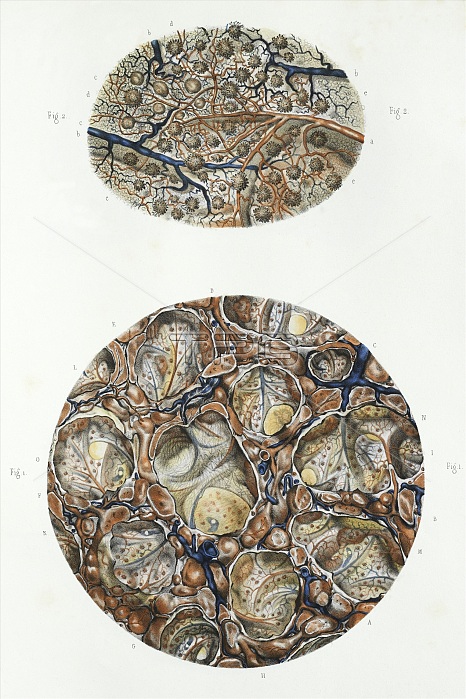
Spleen anatomy. 19th century artwork showing the microscopic structure of the spleen. The upper figure shows the capillaries and lymphatic nodes and vessels. The lower figure shows areas of white pulp within a matrix of red pulp. The red pulp is responsible for filtering blood, while the white plp contains immune system cells that respond to pathogens. This anatomical artwork is plate 45 from volume 5 (1839) of 'Traite complet de l'anatomie de l'homme' (1831-1854). This 8-volume anatomy atlas was produced by the French physician and anatomist Jean-Baptiste Marc Bourgery (1797-1849). The illustrations were by Nicolas-Henri Jacob (1781-1871).
| px | px | dpi | = | cm | x | cm | = | MB |
Details
Creative#:
TOP16632841
Source:
達志影像
Authorization Type:
RM
Release Information:
須由TPG 完整授權
Model Release:
N/A
Property Release:
N/A
Right to Privacy:
No
Same folder images:
 Loading