
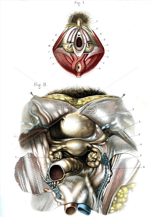
Female genitals. 1866 illustration showing the anatomy of the internal and external female genitals. Fig. 1: vagina (centre), clitoris (upper centre), anus (lower centre) and Bartholin's glands (beige, centre left and right). Fig. 2: ovaries (round, centre left and right), fallopian tubes (curved, centre left and right), uterus (womb, oval centre), bladder (large, oval, upper centre).
| px | px | dpi | = | cm | x | cm | = | MB |
Details
Creative#:
TOP16029574
Source:
達志影像
Authorization Type:
RM
Release Information:
須由TPG 完整授權
Model Release:
N/A
Property Release:
N/A
Right to Privacy:
No
Same folder images:
1800s186619thcenturyanatomicalanatomyartworkbartholin'ssglandbiologicalbiologybladderclitoriscutawayexternalgenitaliafallopiantubefemalegenitalgenitalsgenitourinaryglandsgreatervestibularglandhistoricalhistoryhistoryofsciencehumanbodyillustrationinternalno-onenobodyopeningorganorgansovariesovaryreproductiveorgansecretorysectionsectionedstructurestructurestubesurogenitalsystemvaginavaginalglandvulvavulvovaginalwhitebackgroundwoman
1800s186619thanatomicalanatomyartworkbackgroundbartholin'ssbiologicalbiologybladderbodycenturyclitoriscutawayexternalfallopianfemalegenitalgenitaliagenitalsgenitourinaryglandglandglandglandsgreaterhistoricalhistoryhistoryhumanillustrationinternalno-onenobodyofopeningorganorganorgansovariesovaryreproductivesciencesecretorysectionsectionedstructurestructuressystemtubetubesurogenitalvaginavaginalvestibularvulvavulvovaginalwhitewoman
 Loading