
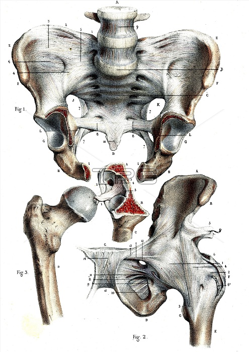
Hip joints. 1866 illustration showing the anatomy of the pelvis and ball-and-socket joints of the hip. Fig. 1: pelvis, including the acetabula (lower left and right), the 'sockets' of the hip joints. Fig. 2: Rear view of the hip joint, showing the tendons. Fig. 3: femur (thigh bone) and femoral head (round, upper right), the 'ball' of the hip joint.
| px | px | dpi | = | cm | x | cm | = | MB |
Details
Creative#:
TOP16029560
Source:
達志影像
Authorization Type:
RM
Release Information:
須由TPG 完整授權
Model Release:
N/A
Property Release:
N/A
Right to Privacy:
No
Same folder images:
1800s186619thcenturyacetabulaacetabulofemoraljointacetabulumanatomicalanatomyartworkballball-and-socketbiologicalbiologybonebonescutoutcutoutscut-outcut-outscutoutcutoutsfemoralheadfemurhiphipshistoricalhistoryhistoryofsciencehumanbodyillustrationjointno-onenobodyorganorganspelvicpelvisskeletalskeletonsocketsocketsstructurestructuressynovialjointtendontendonswhitebackground
1800s186619thacetabulaacetabulofemoralacetabulumanatomicalanatomyartworkbackgroundballball-and-socketbiologicalbiologybodybonebonescenturycutcutcut-outcut-outscutoutcutoutsfemoralfemurheadhiphipshistoricalhistoryhistoryhumanillustrationjointjointjointno-onenobodyoforganorgansoutoutspelvicpelvisscienceskeletalskeletonsocketsocketsstructurestructuressynovialtendontendonswhite
 Loading