
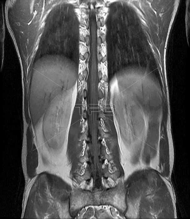
Normal spinal cord. Magnetic resonance imaging scan (MRI) of a section through the torso of a 35-year-old patient, showing the normal appearance of the thoraco-lumbar spinal cord (vertical, centre), with the bulge of the terminal cone and the emergence of the nerve roots to the different lumbar floors.
| px | px | dpi | = | cm | x | cm | = | MB |
Details
Creative#:
TOP16003950
Source:
達志影像
Authorization Type:
RM
Release Information:
須由TPG 完整授權
Model Release:
N/A
Property Release:
N/A
Right to Privacy:
No
Same folder images:
3535-year-old35-years-oldabdomenabdominalanatomicalanatomyblackandwhiteblackbackgroundblack-and-whitecentralnervoussystemcnsdiagnosticimaginghealthyhumanbodylumbarmagneticresonanceimagingmonochromemrinervenervesno-onenobodynormalradiographyradiologicalradiologyrootrootsscansectionsectionedspinalcordspineterminalconethirtiesthoracicthoraco-lumbarthorax
3535-year-old35-years-oldabdomenabdominalanatomicalanatomyandbackgroundblackblackblack-and-whitebodycentralcnsconecorddiagnostichealthyhumanimagingimaginglumbarmagneticmonochromemrinervenervesnervousno-onenobodynormalradiographyradiologicalradiologyresonancerootrootsscansectionsectionedspinalspinesystemterminalthirtiesthoracicthoraco-lumbarthoraxwhite
 Loading