
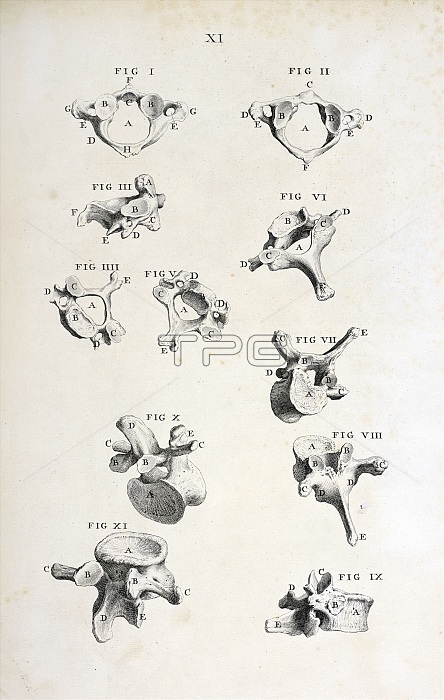
Spinal vertebrae, 18th-century illustration. At top is an atlas vertebra, the uppermost cervical vertebra that joins the neck to the skull. below this is a cervical vertebra, with lumbar vertebrae at centre right and bottom. This illustration is from 'Osteographia, or the Anatomy of the bones' (London, 1733) by English surgeon and anatomist William Cheselden (1688-1752). It was the first full and accurate description of the anatomy of the human skeletal system.
| px | px | dpi | = | cm | x | cm | = | MB |
Details
Creative#:
TOP15985729
Source:
達志影像
Authorization Type:
RM
Release Information:
須由TPG 完整授權
Model Release:
N/A
Property Release:
N/A
Right to Privacy:
No
Same folder images:
1700s173318thcenturyanatomicalanatomyanatomyofthebonesartworkatlasbackboneblack-and-whitebonebonesbookcervicalenglandenglisheuropeanhealthyhistoricalhistoryillustrationlumbarmedicalmedicinemonochromeno-onenobodynormalosteographiaosteologypagepublicationskeletalspinalspinevertebravertebraevertebralwilliamcheselden
1700s173318thanatomicalanatomyanatomyartworkatlasbackboneblack-and-whitebonebonesbonesbookcenturycervicalcheseldenenglandenglisheuropeanhealthyhistoricalhistoryillustrationlumbarmedicalmedicinemonochromeno-onenobodynormalofosteographiaosteologypagepublicationskeletalspinalspinethevertebravertebraevertebralwilliam
 Loading