
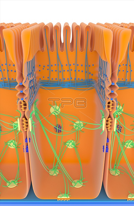
Intestinal cell junctions. Illustration of an intestinal epithelial cell, showing anchoring junctions (desmosomes, yellow; adherens, orange), gap junctions (blue), tight junctions (ridged) and a hemidesmosome junction (bottom). Cytoskeletal filaments are blue and green. Across top are the microvilli that absorb nutrients from the intestinal lumen as food is digested. The various junctions shown here have a range of functions. Tight junctions form a relatively impermeable barrier. Anchoring junctions provide mechanical support. Gap junctions allows chemical and electrical communication between cells. For this artwork with labels, see C023/8827.
| px | px | dpi | = | cm | x | cm | = | MB |
Details
Creative#:
TOP15984359
Source:
達志影像
Authorization Type:
RM
Release Information:
須由TPG 完整授權
Model Release:
N/A
Property Release:
N/A
Right to Privacy:
No
Same folder images:
 Loading