
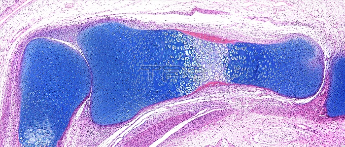
Light microscopy of a fetal finger bone. At this time of development the future bone is mostly cartilage (blue tissue) with a primary ossification centre in the middle. Here the cartilage cells have enlarged prior to their programmed death that will leave behind a cartilage matrix upon which new bone matrix will be deposited by osteoblasts. A collar of early bone stained red is seen at this central location. Over time most of the cartilage will be replaced with bone leaving a cap of articular cartilage covering the bone ends. This type of bone formation is called endochondral ossification and occurs in almost all of the bones of the axial skeleton and limbs. Magnification x85 when narrow width printed at 10 cm.
| px | px | dpi | = | cm | x | cm | = | MB |
Details
Creative#:
TOP15205088
Source:
達志影像
Authorization Type:
RM
Release Information:
須由TPG 完整授權
Model Release:
No
Property Release:
No
Right to Privacy:
No
Same folder images:
 Loading