
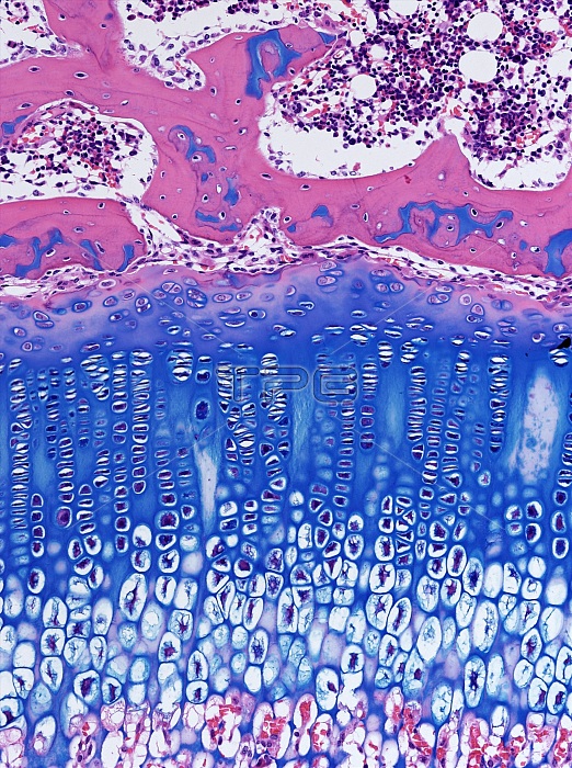
Light microscopy of cellular types of a growth plate in a growing bone. The growth plate is located towards the end of the bone at the epiphysis. It is formed of cartilage consisting of columns of cartilage cells (chondrocytes) and the matrix which they produce (deep blue). Mature chondrocytes enlarge then shrink just prior to cell death leaving empty shells of cartilage matrix. This cartilage persists and acts as a surface upon which bone-forming cells (osteoblasts) arrive in blood vessels (pink) in the lower aspect of the image. Osteoblasts deposit new bone on the cartilage scaffolds by a process termed endochondral ossification. Over time the bone slowly grows in length until the growth plate stops making cartilage. Magnification x180 when narrow
| px | px | dpi | = | cm | x | cm | = | MB |
Details
Creative#:
TOP15070399
Source:
達志影像
Authorization Type:
RM
Release Information:
須由TPG 完整授權
Model Release:
No
Property Release:
No
Right to Privacy:
No
Same folder images:
 Loading