
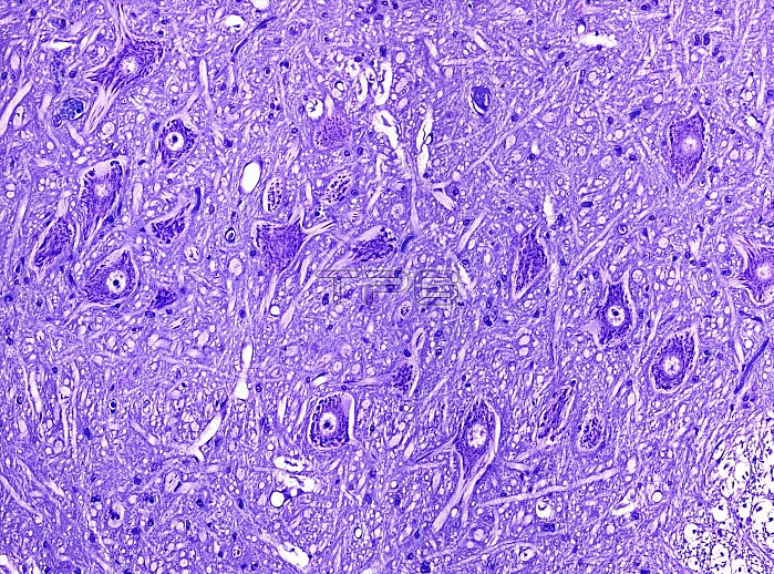
Light microscopy of motor neurons in spinal cord grey matter. The cell bodies of motor neurons show a central nucleus and peripheral cytoplasm (deep purple). In histologic sections processes of dendrites and an axon extend from each cell body. In humans one such axon may be a metre in length if supplying the foot. Tissue between the cell bodies is called the neuropil and is formed of intermingling dendrites, axons, capillaries and glial cells. Magnification x100 when narrow width printed at 10 cm.
| px | px | dpi | = | cm | x | cm | = | MB |
Details
Creative#:
TOP15070390
Source:
達志影像
Authorization Type:
RM
Release Information:
須由TPG 完整授權
Model Release:
No
Property Release:
No
Right to Privacy:
No
Same folder images:
 Loading