
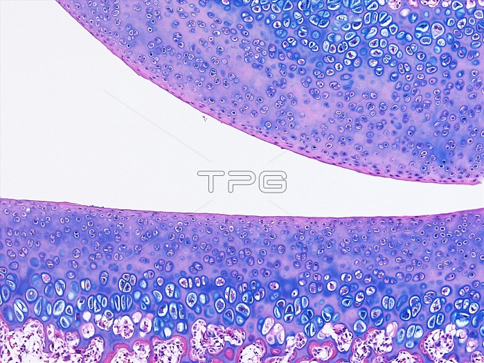
Light microscopy of cartilage covering the surfaces of two bone ends inside a joint. Called hyaline articular cartilage it consists of a matrix and cartilage cells (chondrocytes) stained blue. Some functions of this cartilage are to provide an almost frictionless surface for joint movement and, by retaining water in its matrix, to resist compression. The joint space is filled with synovial fluid acting as a lubricant and nutrient source. The cartilage has no direct blood vessels. Magnification x120 when narrow width printed at 10 cm.
| px | px | dpi | = | cm | x | cm | = | MB |
Details
Creative#:
TOP15070384
Source:
達志影像
Authorization Type:
RM
Release Information:
須由TPG 完整授權
Model Release:
No
Property Release:
No
Right to Privacy:
No
Same folder images:
 Loading