
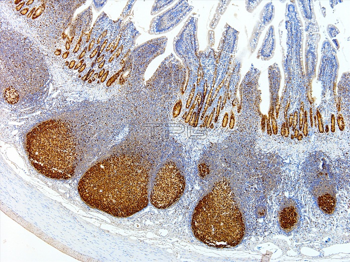
Light microscopy of lymphoid follicles in a Peyer's patch of the ileum, the last segment of the small bowel. The lymphoid cells are stained brown with a immunostain for any cells that are about to divide or are proliferating. Dividing cells in the intestinal crypts are similarly stained. The follicles supply lymphocytes to the local tissue for immune defence against harmful antigens such as toxins and infectious micro-organisms. Peyer's patches and lymphoid components of the intestines are collectively referred to as gut-associated lymphoid tissue abbreviated as GALT. Magnification x40 when printed at 10 cm.
| px | px | dpi | = | cm | x | cm | = | MB |
Details
Creative#:
TOP14988141
Source:
達志影像
Authorization Type:
RM
Release Information:
須由TPG 完整授權
Model Release:
N/A
Property Release:
No
Right to Privacy:
No
Same folder images:
 Loading